Yesterday was our fetal echo follow-up appointment. Even though everything looked good at our first appointment, we were advised in January to return for a second look around this time in the pregnancy, just to ensure everything was growing and forming normally.
Just like last time, a technician took a bunch of measurements over the course of about 15 minutes and then left the room to show them to the doctor. I didn't have a view of the monitor this time around and Shep didn't want to look at the monitor because he didn't want to mistakenly see any "below the waist" body parts that might ruin our big surprise, so I stared at the ceiling hoping everything was okay while he played Words With Friends on his phone.
Unlike last time, before the technician left the room she asked me to continue laying on the table with the ultrasound goop on my belly in case the doctor needed to take a closer look. The paranoid part of me started to get a teensy bit worried at that request -- did that mean something was wrong?? When Dr. Phoon came in a few minutes later he explained that 2 of the measurements the technician had taken were not consistent with one another, and that although both numbers fell within the normal range, they were each on opposite sides of that range. Hmmm... So, he wanted to take a look himself just to ensure everything measured up as it should.
Now for the GREAT news...
According to Dr. Phoon, the aortic arch on our baby looks "perfect" and there is no evidence to indicate any obstruction. Apparently all of the valves are formed early, and then they grow throughout the pregnancy. Per the doctor, all of the structures of our baby's heart are formed "perfectly" such that even if the baby was born today, the heart would work fine for a newborn. Phew, talk about a relief!!!
Approximately 1-2% of the population has a bicuspid aortic valve. However, NYU does over 5,000 pediatric echocardiograms a year and they have not seen bicuspid aortic valves in 1-2% of those children, nor does the occurrence rate in children generally match that of the general population, which leads many researchers and doctors across the world to believe that the condition can develop over the course of one's lifetime. This certainly seems to make sense, since many people are not diagnosed with this condition until adulthood. For this reason, the doctor recommended we consider getting our child's heart checked out sometime before school begins, perhaps around 3-4 years old, and to have a low threshold for getting the heart checked out before that time if at any point a pediatrician discovers a murmur in our baby's heart. But, as long as everything appears fine when the baby is born, and the baby seems generally healthy, there's no reason to be concerned or to consult a pediatric cardiologist upon birth!
The doctor did mention that altough occurrence rates are 10-15% higher if a parent has a bicuspid aortic valve, that still eaves an 85% chance for a normal, healthy heart, so there's plenty of reason to be optimistic that this will be our last visit to the Pediatric & Fetal Echocardiography Lab. :)
Showing posts with label congenital heart defects. Show all posts
Showing posts with label congenital heart defects. Show all posts
Wednesday, March 12, 2014
Tuesday, January 7, 2014
Fetal Echo Appt
Today was our Fetal Echocardiogram appointment and, much to my relief, it went well! A fetal echo is a detailed scan of a baby's heart to look for congenital heart defects. We underwent this non-invasive procedure due to an aortic valve defect on Hubby's side, which can be inherited. The purpose of the exam was to determine whether any heart issues are currently presenting in our baby, and if so, what treatment options/risks would be.
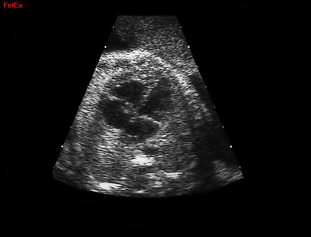
Here's a sample view of a normal fetal heart, courtesy of the UPenn Medical Library:
The echo portion took about 20 minutes, during which the technician did lots of measuring and took photos at different angles using what looked and worked very much like a regular ultrasound wand. We also heard the heart beating, which for me is always a huge relief. I was pretty impressed that the technician was able to get all the info he needed relatively quickly despite the fact that baby kept flipping and darting around the whole time! It appears we have a "very active baby" on our hands -- wonder where baby got those genes from?! Certainly not me... ;) Despite our baby's heart measuring in at a mere 1.5 cm {which is normal, but so small to think about!}, the technician was able to identify clearly the four chambers of the heart and all appropriate major valves and vessels. Phew!
After the technician performed the echo we met with a pediatric cardiologist to review the results. The doctor advised us that everything looked good, and that although it is too soon to spot a bicuspid aortic valve at this point, in most cases a healthy 20 week scan means the baby will be born with a healthy heart because most of the time {I think he mentioned an estimate of 80%} left heart defects manifest themselves by 20 weeks. The inheritance rate for left heart lesions is currently estimated in the 10-20% range, while other heart defects are typically inherited only ~ 3-5% of the time. Left heart lesions can evolve before birth though, so as a precaution, he recommended that we return for a follow-up echo in 8-10 weeks to ensure that everything has grown appropriately and the aortic arch looks good. If they spot something odd at the follow-up appointment, they'll have a specialist examine the baby at the hospital after delivery. But, if all looks good at the follow-up appointment -- which is what we're hoping for -- then there's likely no need for any further concern and we'd just get a pediatric cardiologist to examine the child sometime within the first few years of life just to triple check everything.
So, for the time being we have every reason to hope for the best and try not to worry! :) Though I'm starting to realize that as a soon-to-be parent that is much easier said than done!!
As a side note, both the sonogram technician and the doctor were incredibly nice and explained everything clearly. Despite being at a top medical facility with lots of patients, they each entertained all questions we had, and did not rush us at all. We also were not kept waiting any longer than a few minutes beyond our original appointment start time. This is all quite a novelty in NYC!
If you're interested, you can read more about Fetal Echocardiograms here.
Here, you can find some facts and information on congenital heart disease and pediatric heart conditions.
Here's a sample view of a normal fetal heart, courtesy of the UPenn Medical Library:
 |
| Source |
After the technician performed the echo we met with a pediatric cardiologist to review the results. The doctor advised us that everything looked good, and that although it is too soon to spot a bicuspid aortic valve at this point, in most cases a healthy 20 week scan means the baby will be born with a healthy heart because most of the time {I think he mentioned an estimate of 80%} left heart defects manifest themselves by 20 weeks. The inheritance rate for left heart lesions is currently estimated in the 10-20% range, while other heart defects are typically inherited only ~ 3-5% of the time. Left heart lesions can evolve before birth though, so as a precaution, he recommended that we return for a follow-up echo in 8-10 weeks to ensure that everything has grown appropriately and the aortic arch looks good. If they spot something odd at the follow-up appointment, they'll have a specialist examine the baby at the hospital after delivery. But, if all looks good at the follow-up appointment -- which is what we're hoping for -- then there's likely no need for any further concern and we'd just get a pediatric cardiologist to examine the child sometime within the first few years of life just to triple check everything.
So, for the time being we have every reason to hope for the best and try not to worry! :) Though I'm starting to realize that as a soon-to-be parent that is much easier said than done!!
As a side note, both the sonogram technician and the doctor were incredibly nice and explained everything clearly. Despite being at a top medical facility with lots of patients, they each entertained all questions we had, and did not rush us at all. We also were not kept waiting any longer than a few minutes beyond our original appointment start time. This is all quite a novelty in NYC!
If you're interested, you can read more about Fetal Echocardiograms here.
Here, you can find some facts and information on congenital heart disease and pediatric heart conditions.
 |
| Source |
Tuesday, November 12, 2013
Nuchal Translucency Screening
{Pardon the tardiness on this topic -- I started this post months ago but never finished it. Better late than never though, right?!}
The nuchal translucency scan takes place in the latter part of the first trimester, sometime during weeks 11-13. It's a special ultrasound scan designed to screen for chromosomal defects -- particularly trisomy 13, 18 and 21 {Down Syndrome}, in addition to congenital heart defects.
Combined with specific blood tests, the nuchal translucency ultrasound is called the "first trimester screening". It's a "non-invasive" way of assessing a baby's risk of chromosomal abnormalities, which means there are no needles and no physical risk to having the screening done. Since this test is a screening {as opposed to a diagnostic test} it will not provide a 100% accurate or definitive answer -- it's more of a gauge of whether things look generally normal or problematic. It gives the doctor a general indication of whether there is cause for concern, or need for additional testing, such as an amniocentesis or CVS test.
Here's an illustration of the measurement the ultrasound is designed to screen, along with a more detailed explanation of the screening taken from this source:
Our NT screening took place on November 7th, at 11.5 weeks gestation. I have to admit, I was pretty nervous going into it, especially since we knew our first baby's miscarriage was a result of chromosomal abnormalities. The 20 minutes or so spent in the office waiting room before being called in seemed to crawl by {especially since I had to pee but wasn't allowed}, and the several minutes during the screening during which the technician measured and took photos and notes but didn't provide any sort of medical insight lasted FOREVER.
The technician did, however, call our baby "cute" while she was conducting her measurements. While I'm sure she says that to all the patients, we took it to heart and totally agree. :)
After about 10-15 minutes of ultrasound measurements and photos, we were escorted in to meet with the doctor. Thankfully, she had EXCELLENT news for us. According to the screening, our chances of having a baby with any chromosomal abnormalities were extremely low. Our specific risk profile for Down Syndrome is 1 in 4,463, and for Trisomy 18/13 is 1 in >10,000. Better yet, the risk ratios matched that of a 20 year old mother, which made me feel pretty good since I'll be turning 33 in a couple weeks. ;) Needless to say, we both breathed a HUGE sigh of relief after receiving these results. Though there are no guarantees, these odds are pretty favorable!
The NT is often followed up with a second trimester blood draw {usually called a multiple marker, triple or quad screen} to check on the probability of open neural tube defects. We'll definitely be waiting for those results, but for now will just be thankful for these great numbers!
The nuchal translucency scan takes place in the latter part of the first trimester, sometime during weeks 11-13. It's a special ultrasound scan designed to screen for chromosomal defects -- particularly trisomy 13, 18 and 21 {Down Syndrome}, in addition to congenital heart defects.
Combined with specific blood tests, the nuchal translucency ultrasound is called the "first trimester screening". It's a "non-invasive" way of assessing a baby's risk of chromosomal abnormalities, which means there are no needles and no physical risk to having the screening done. Since this test is a screening {as opposed to a diagnostic test} it will not provide a 100% accurate or definitive answer -- it's more of a gauge of whether things look generally normal or problematic. It gives the doctor a general indication of whether there is cause for concern, or need for additional testing, such as an amniocentesis or CVS test.
Here's an illustration of the measurement the ultrasound is designed to screen, along with a more detailed explanation of the screening taken from this source:
| Source |
The nuchal translucency is the fluid found at the back of your baby’s head and neck, just beneath the skin. The thickness of this fluid can be precisely measured and this is called the nuchal translucency (or NT) measurement. Normally the amount of fluid is small, producing a thin NT measurement.
We know that the amount of fluid can increase in the presence of certain conditions, producing a thicker NT measurement. Conditions associated with an increased NT measurement include some chromosome abnormalities (for example, trisomy 13, 18 and 21) and some structural problems (for example, heart abnormalities). An increased NT measurement does not always mean the baby has a problem but it does increase the risk.
{You can also read more about the NT test here and here.}
Our NT screening took place on November 7th, at 11.5 weeks gestation. I have to admit, I was pretty nervous going into it, especially since we knew our first baby's miscarriage was a result of chromosomal abnormalities. The 20 minutes or so spent in the office waiting room before being called in seemed to crawl by {especially since I had to pee but wasn't allowed}, and the several minutes during the screening during which the technician measured and took photos and notes but didn't provide any sort of medical insight lasted FOREVER.
The technician did, however, call our baby "cute" while she was conducting her measurements. While I'm sure she says that to all the patients, we took it to heart and totally agree. :)
After about 10-15 minutes of ultrasound measurements and photos, we were escorted in to meet with the doctor. Thankfully, she had EXCELLENT news for us. According to the screening, our chances of having a baby with any chromosomal abnormalities were extremely low. Our specific risk profile for Down Syndrome is 1 in 4,463, and for Trisomy 18/13 is 1 in >10,000. Better yet, the risk ratios matched that of a 20 year old mother, which made me feel pretty good since I'll be turning 33 in a couple weeks. ;) Needless to say, we both breathed a HUGE sigh of relief after receiving these results. Though there are no guarantees, these odds are pretty favorable!
The NT is often followed up with a second trimester blood draw {usually called a multiple marker, triple or quad screen} to check on the probability of open neural tube defects. We'll definitely be waiting for those results, but for now will just be thankful for these great numbers!
Subscribe to:
Posts (Atom)
